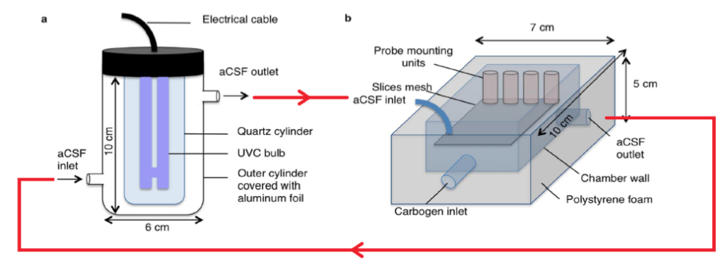
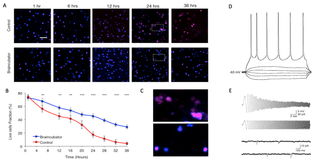
Slice Viability – Standard Control v’s Braincubator
A) Confocal microscopic serial images (×40) of acute brain slices following different incubation time at contol and Braincubator conditions. All sections were sliced at the same time. The combination of DAPI (blue) and PI (red) allows the simultaneous visualisation of all cells in the slice. Scale bar = 50 μm. B) Graph depicting the slice viability as assessed from the cell death/total assay. For each time point n = 6, data displayed as Mean ± S.E.M. (C) Expansion of the marked areas in A depicting the morphology of dead cells following 24 hours in control (Top) and Braincubator (Bottom).(D) I-V traces. Increasing step currents of 500 ms were injected into the soma through the recording electrode to reveal the input resistance and firing properties. (E) The resonance frequency was measured by injecting a chirp stimulation of 60 pA (peak to peak). Sample traces of spontaneous synaptic activity recorded from a neuron after 31 hrs in the Braincubator.